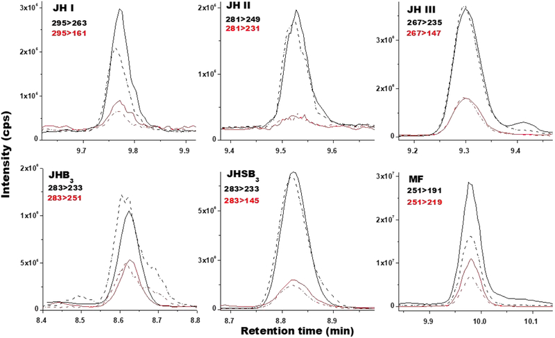
Fig. 4: Analysis of JH homologs from biological samples:
Typical ion extracted chromatograms comparing the relationships between retention times in minutes (X-axis) and signal intensities (cps; counts per second) (Y-axis) for JHs from biological samples (solid line) and from standard solutions (dashed lines). MRM transitions: primary (black) and secondary (red). JH I and II are from B. mori larval hemolymph. JH III is from Ae. aegypti adult female hemolymph. JHB3 is from D. melanogaster larval hemolymph. JHSB3 is from Dipetalogaster maxima adult female hemolymph. MF was synthesized in vitro by the CA of Ae. aegypti 4th instar larvae. Primary transitions masses evaluated are in black and secondary transitions are in red.

