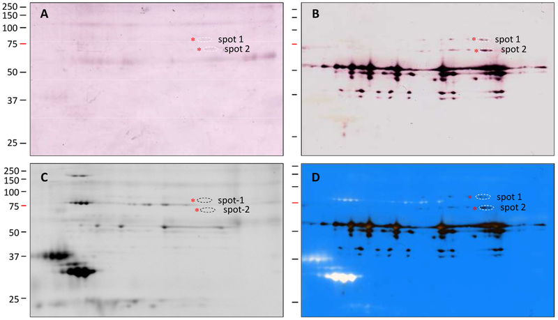
Fig. 3.
Separation of the hemolymph proteins eluted from the serpin-12 antibody column by 2DE for immunoblot and LC-MS/MS analysis. As described in Section 2.3, an induced plasma sample was treated with S. aureus peptidoglycan and then separated by serpin-12 immunoaffinity chromatography. The eluted proteins (200 μg) were resolved by 2DE followed by immunoblot analysis using diluted antiserum to HP14PD (A, 1:1000) or serpin-12 (B, 1:2000), by staining with SYPRO Ruby (C), or by superposition (D) of panels B and C. After image alignment, spot-1 and spot-2, marked by dashed ovals, were excised from the stained gel for trypsin digestion and mass spectrometric analysis. Positions and sizes (in kDa) of the pre-stained Mr standards are marked on the left, with the 75 kDa marker highlighted red.

