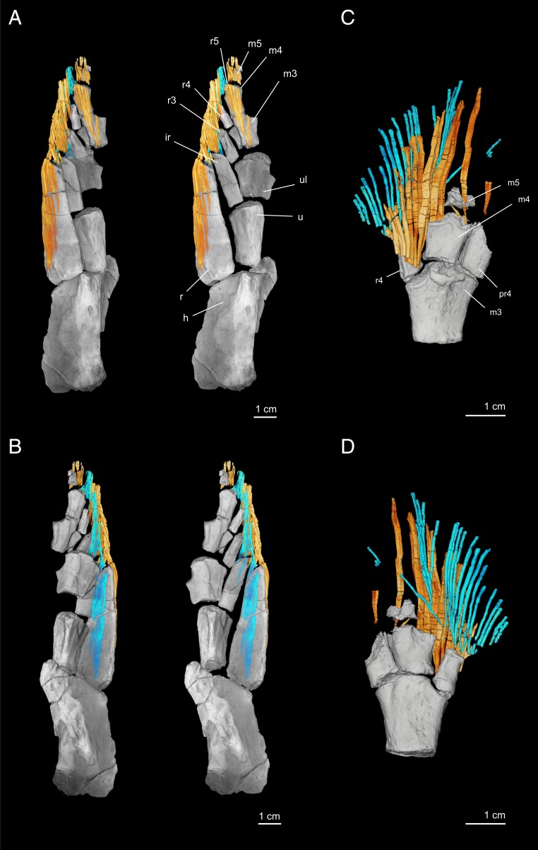
Fig. 3.
Dermal rays of the pectoral fin of T. roseae. NUFV110 in (A) dorsal and (B) ventral perspectives. A, Left and B, Left show the specimen in the preserved position with scales and matrix removed. A, Right and B, Right show endoskeletal elements repositioned to more natural, articulated positions. NUFV109 in (C) dorsal and (D) ventral perspectives. In all images, dorsal hemitrichia are shown in yellow–orange, and ventral hemitrichia are cyan. h, humerus; ir, intermedium; m3, third mesomere; m4, fourth mesomere; m5, fifth mesomere; pr4, posterior radial adjacent to mesomere 4; r, radius; r3, radial of the third mesomere; r4, radial of the fourth mesomere; r5, radial of the fifth mesomere; u, ulna; ul, ulnare.

