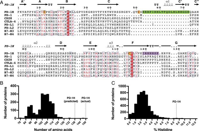
Fig. 1.
PD-1H has unique sequence features in the CC′ and FG loops, a very long IgV domain, and a large number of conserved histidines. (A) Multiple-sequence alignment of PD-1H and CD28/B7 family member IgV domains. Secondary structural annotations are based on our crystal structure of PD-1H. Similar residues are colored red, and invariant residues have red backgrounds. A long insertion in PD-1H between the C- and C′-strands is highlighted in green, the PD-1H sequence corresponding to the CDR3-like region in the FG loop is highlighted in purple, and additional cysteines that are invariant among PD-1H orthologs are highlighted in orange. (B) Histogram of all IgV domain lengths as defined by Pfam. (C) Histogram of histidine content in all annotated type I transmembrane ECDs.

