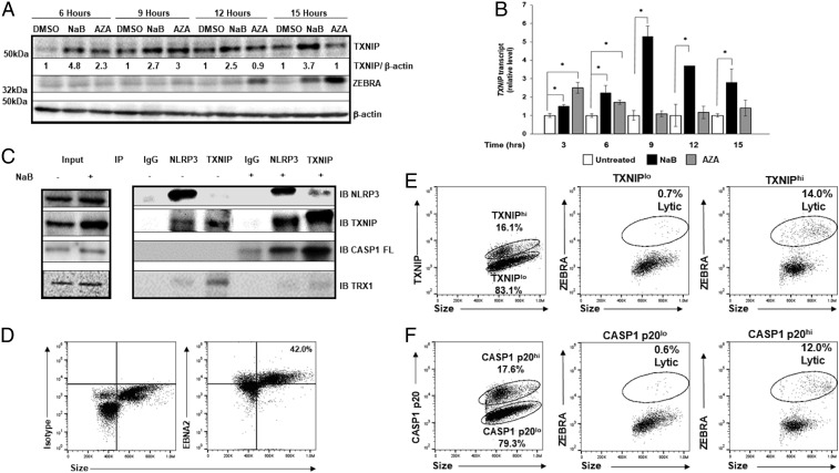
Fig. 3.
Early increase in TXNIP and activation of the NLRP3 inflammasome precedes expression of the replication switch. (A and B) HH514-16 cells were exposed to lytic triggers NaB and 5-AZA-2′-deoxycytidine (versus dimethyl sulfoxide [DMSO] control) and harvested at indicated time points for immunoblotting with (A) indicated antibodies or (B) qRT-PCR. Numbers in A represent ratios between indicated proteins. Experiments were performed at least three times, and data in B represent averages of three independent experiments; error bars, SEM; *P ≤ 0.05. (C) HH514-16 cells were treated with NaB (versus untreated control) for 6 h, cell lysates were immunoprecipitated using indicated antibodies (IP), and bound proteins were detected by immunoblotting with indicated antibodies (IB). Input lysates are shown on the left. (D–F) CD19+ B cells from the blood of a patient with PTLD were analyzed by flow cytometry for EBNA2 and size to demarcate EBNA2+ “blast-like” large cells for further analysis; number indicates percent EBNA2+ cells (D). EBV-infected B blast-like cells were analyzed by flow cytometry for TXNIP (E) or cleaved caspase-1 (CASP1 p20; F) expression (Left dot plots), and percent TXNIPhi versus percent TXNIPlo cells (E) or percent CASP1 p20hi versus percent CASP1 p20lo cells (F) expressing the lytic switch protein ZEBRA are shown (Right dot plots). Data from a representative of 3 patients is shown.

