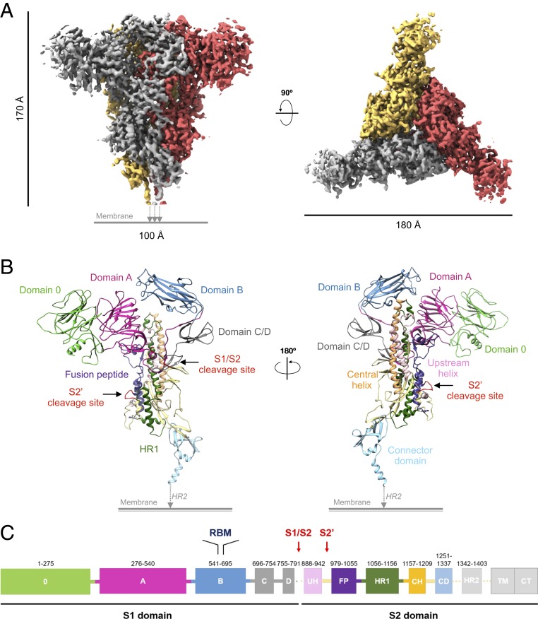
Fig. 1.
Cryo-EM structure of FIPV-UU4 S protein. (A) The 3.3-Å cryo-EM map of FIPV-UU4 S protein shown in side view (Left) and top view (Right) with the 3 protomers colored in gold, red, and gray. (B) Cartoons representative of the atomic model of monomeric FIPV-UU4 S protein. (C) Functional subunits and domains are indicated and colored as defined in the schematic representation as a function of the sequence number indicated above each functional unit. CD, connector domain; CH, central helix; CT, cytoplasmic tail; FP, fusion peptide; TM, transmembrane domain; UH, upstream helix. Regions that were resolved by cryo-EM, namely HR2, TM, and CT, are shown in light gray.

