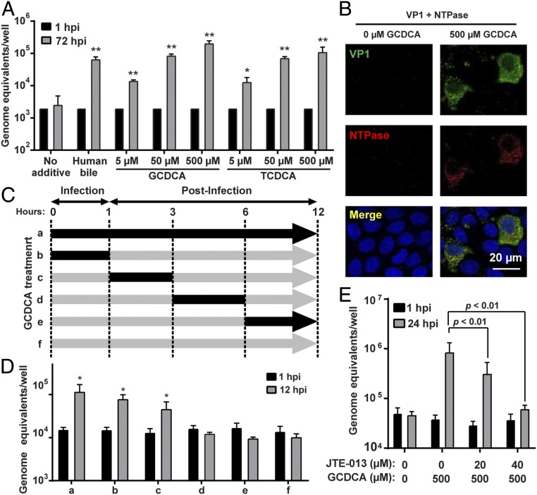
Fig. 2.
BAs are required early in GII.3 infection. (A) HIE monolayers were infected as in Fig. 1 with GII.3 in the presence or absence of GCDCA and TCDCA for 72 h. *P < 0.05 and **P < 0.01 comparing GEs at 72 hpi to 1 hpi. (B) HIE monolayers infected with 4.3 × 10⁶ GEs in the presence or absence of 500 µM GCDCA. VP1 and NTPase were detected by confocal laser-scanning microscopy using guinea pig anti-GII.3 VLP (green) and rabbit anti-GII.3 NTPase (red) antisera. Nuclei were stained with DAPI (blue). (Scale bar, 20 µm.) (C) Schematic showing with black arrows when 500 µM GCDCA was added to the medium during GII.3 infection of HIEs. (D) HIE monolayers were infected with GII.3 for 12 h. GCDCA was added to the medium as in C. HIEs were washed three times with CMGF(−) at the end of each period. *P < 0.05 comparing GEs at 12 hpi to 1 hpi. (E) S1PR2 antagonist, JTE-013, and GCDCA were added to the medium at the indicated concentrations and infected with GII.3 as in Fig. 1 for 24 h. P values between conditions are indicated.

