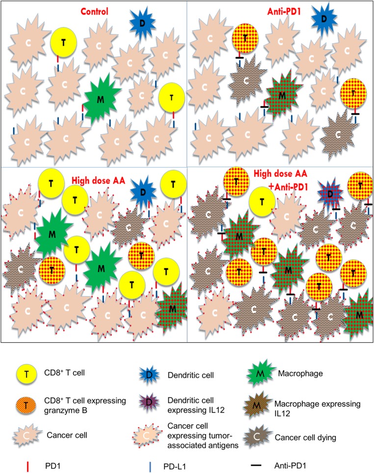
Fig. 7.
Graphical summary of the synergistic effects of high-dose AA and anti-PD1. Single-agent anti-PD1: blocks the inhibitory PD-1/PD-L1 axis, thereby activating the sparse infiltrated cytotoxic T lymphocytes, NK cells (not depicted), and macrophages. Single-agent high-dose AA: enhances tumor immune recognition and increases macrophage and cytotoxic T cell infiltration into the tumor. However, despite the increased immune cell numbers, the inhibitory effect of PD-1/PDL-1 axis on cytotoxic cells and macrophages cannot be abrogated by AA. Anti-PD1+high-dose AA: The combination treatment results in both increased immune infiltration and enhanced activation of APCs and cytotoxic cells, resulting in marked tumor shrinkage. Red dots within the T cells represent granzyme B. Red dots within the macrophages and dendritic cells represent IL-12, which activates the cytotoxic cells (cytotoxic T cells and NK cells). Red dots on the surface of cancer cells represent increased HERV expression with AA treatment (or other potential immune-recognition enhancing antigenic epitopes). Black dots within the cancer cells represent dying cells. Red mark on T cells and macrophages represents PD1. Blue mark on cancer cells represents PDL1. Black mark between the red and blue marks represents anti-PD1 antibody.

