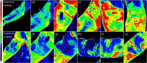
Fig. 5.
Examples of perfusion in scalds with various healing times in two different children. On the upper row, a wound on the right upper and lower arm is shown that contains regions that healed between 9 and 17 days. Perfusion images (a)–(e) are acquired at (a) 14 h, (b) 4 days, (c) 6 days, (d) 8 days, and (e) 15 days after the injury. On the lower row, a wound on the upper arm of another patient is shown, which contains different regions that did not heal after 14 days and subsequently underwent surgery. Perfusion images (f)–(j) are acquired at (f) 5 h, (g) 3 days, (h) 6 days, (i) 8 days, and (j) 10 days after the injury. The asterisks (*) indicates area with erroneously low perfusion values due to specular reflections. The color bar on the left side indicates the perfusion scale (0 to 500 PU). Figure reprinted from Fig. 3 in Ref. 52, © 2019, with permission from Elsevier.

