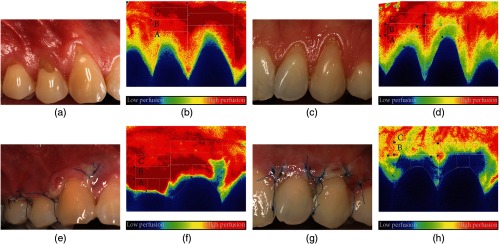
Fig. 9.
Representative photographs and LSCI images of (a), (b), (e), and (f) male and (c), (d), (g), and (h) female subjects; in both cases, a combination of the modified coronally advanced tunnel and Geistlich Mucograft was used. (a)–(d) Images representing the preoperative perfusion and (e)–(h) images showing the wound healing and perfusion 3 days postoperatively. Capital letters (A, B, and C) indicate the regions of interest for the blood flow evaluation. Figure reprinted from Fig. 1 in Ref. 100, © 2019, with permission from Hindawi.

