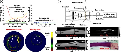
Fig. 8.
Multispectral pulse-based IV-PA image examples that were generated by (a) interpreting spectral variation of different plaque constituents and (b) employing an arithmetic subtraction algorithm. P1, healthy artery region; P2, lipid-rich plaque region. Compared to the less-complex single-wavelength counterpart, multispectral approaches in general improved imaging specificity toward lipids. Reproduced with permission from Refs. 83 and 84.

