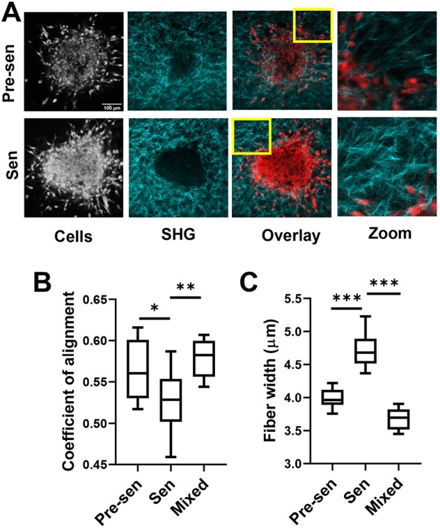
Fig. 6.

Senescent MSCs rapidly remodeled the extracellular matrix. (A) Spheroids formed from BCCs and MSCs [pre-senescent (Pre-sen), senescent (Sen)] embedded in collagen gels were imaged by multiphoton microscopy using SHG to identify collagen (shown in cyan) and for NucRed fluorescence to identify cells. (B,C) SHG images were analyzed using CurveAlign to quantify collagen fiber properties and orientation. Coefficient of alignment measuring degree of collagen alignment surrounding spheroids was enhanced with pre-senescent cells (B), whereas collagen fiber width was increased for spheroids containing senescent cells only (C). Data are shown as box-and-whisker plots, where the box represents the 25–75th percentiles, and the median is indicated. The whiskers show the minimum and maximum value of the data points. *P<0.05, **P<0.01, ***P<0.001 (Student's t-tests).
