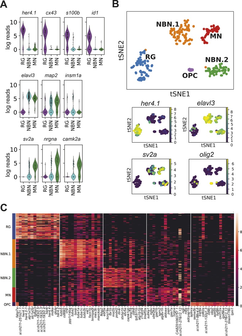
Fig. 3.

Single cell RNA-seq reveals the diversity of radial glia progeny in the adult forebrain. (A) Violin plots showing the expression of known radial glia markers (top), pan-neuronal markers (middle, elavl3 and map2), an early neurogenic fate marker (middle, insm1a) and markers of mature synaptically integrated neurons (bottom). (B) tSNE plot revealing five different clusters from a total of 264 RG, NBN and MN cells (top). The cell number per cluster is: RG, 76 cells; NBN.1, 80 cells; NBN.2, 54 cells; MN, 44 cells; and OPC, 10 cells. Smaller panels underneath show expression of characteristic marker genes that separate the different clusters. (C) Heat map with the top 15 cluster-specific genes for the five identified clusters.
