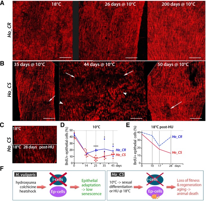
Fig. 3.
Disorganization of the epithelial epidermal layer in aging Ho_CS animals. (A,B) Phalloidin staining of the epidermis in Ho_CR and Ho_CS animals transferred to 10°C and fixed at the indicated time points. Arrows indicate disorganized regions of the epidermis, arrowheads indicate shortened myofibrils. Scale bars: 50 µm. (C) Phalloidin staining of the epidermis in control or HU-treated Ho_CS animals maintained at 18°C and fixed after 28 days. (D,E) BrdU-labeling index values measured after 96 h BrdU exposure performed at the indicated time points either after transfer to 10°C (D) or after HU treatment at 18°C as in Fig. 2E. In D, each dot corresponds to a replicate in which at least 300 cells were counted. In E, animals were maintained at 18°C. *P<0.05, ****P<0.0001 (unpaired t-test). (F) Scheme comparing the impact of i-cell loss in Hv animals, in which epithelial stem cells adapt (Wenger et al., 2016), and in Ho_CS, in which a more limited i-cell loss is lethal, suggesting a lack of epithelial adaptation.

