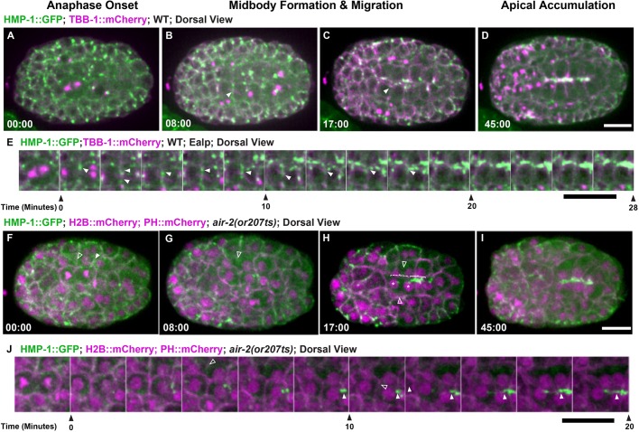
Fig. 4.
Aurora B is required for adhesion dynamics during E8-E16 cytokinesis. Adhesion dynamics during E8-E16 division and polarization. (A-D) HMP-1::GFP (green; microtubules in magenta) localizes to the furrow and midbody (B, arrowhead) during cytokinesis. HMP-1::GFP migrates with the midbody (C, arrowhead) to the apical surface where it accumulates after polarization (D). (E) Montage of HMP-1::GFP during E8-E16 division shows furrow and midbody (arrowheads) migration to the apical midline. (F-I) Aurora B mutants have reduced HMP-1::GFP on the furrow and midbody (F, unfilled arrowheads). When cells fail cytokinesis (G,H, unfilled arrowheads), HMP-1 accumulation is delayed (dashed bracket shows failed cytokinesis, solid bracket indicates successful E8 division with apical accumulation). Asterisks in H indicate nuclei. (I) HMP-1 signal eventually spreads along the midline. (J) Montage of HMP-1::GFP in Aurora B mutant E8-E16 cells that fail cytokinesis (unfilled arrowheads indicate furrow regression) and have delayed apical accumulation. Filled arrowheads indicate HMP-1 signal. Scale bars: 10 μm.

