Fig. 5.
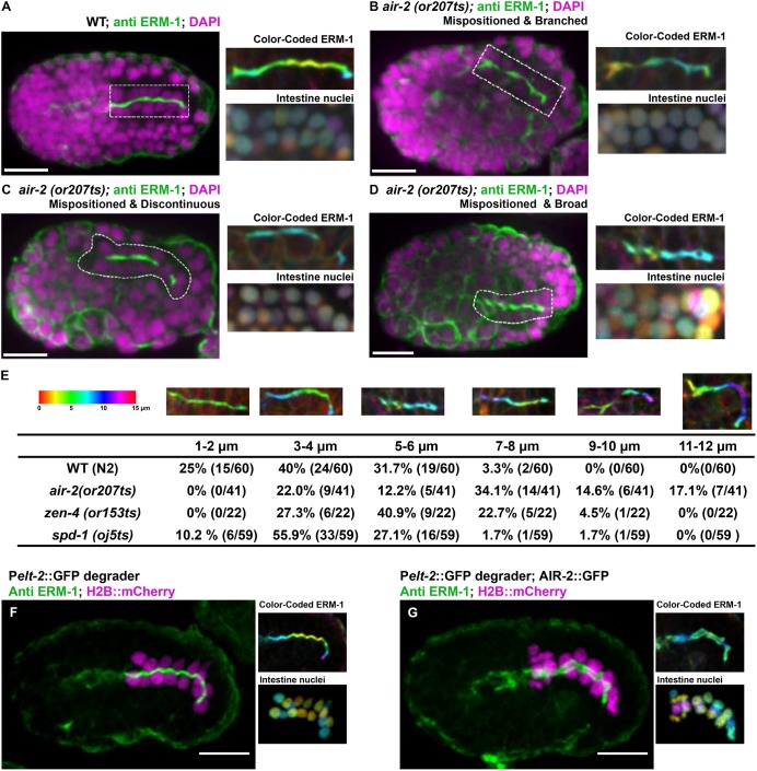
Gut morphogenesis is disrupted in cytokinesis mutants. Apical surface staining after E8-E16 division and polarization. (A) ERM-1 apical staining (dashed rectangle) in wild-type (WT) bean-stage embryos. Maximum z-projected images of ERM-1 and nuclei color-coded according to z-depth (scale shown in F) show tissue organization. (B-D) In air-2(or207) embryos, apical surfaces are mispositioned (B-D), branched (B), contain gaps (C) or have broader staining (D). (E) Quantification of the defective apical z-plane distribution in different mutants (more colors indicate greater distortion in the z-plane). (F) ERM-1 staining and distribution of nuclei in a control embryo (GFP degrader only expressed) shows normal lumen width (1.15±0.11 µm, n=10) and nuclear distribution. (G) Endogenous AIR-2::GFP degradation in the intestine results in significantly broadened ERM-1 staining (2.53±0.33 µm, n=8) and disorganized nuclei. Scale bars: 10 μm.

