Fig. 6.
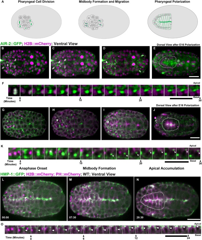
Cytokinesis during pharyngeal precursor cell polarization. (A) Illustration of cell division in PPCs with Aurora B (green; midbody ring in magenta). (B-E) PPC division labeled with AIR-2::GFP (green; H2B::mCherry in magenta) from both ventral (B-D, dashed line highlights one cell, arrowhead indicates midbody) and dorsal (E, dashed line highlights pharynx) views. AIR-2::GFP localizes to chromosomes in metaphase (B), moves to the central spindle in anaphase (C), and appears on the midbody which moves toward the midline (D). AIR-2 persists at the pharyngeal apical surface for an extended time (E). (F) Montage showing AIR-2::GFP migrating toward the midline. (G-K) Imaging of NMY-2::GFP (green; TBB-1::mCherry in magenta), during midbody migration to the midline (I,K). NMY-2::GFP accumulates at the midline during apical constriction (J). (L-N) During PPC cytokinesis, α-catenin (HMP-1::GFP, green; tubulin in magenta) accumulates on the furrow (arrowhead in L) and adjacent to the midbody (arrowhead, M) before accumulating at the midline (N). (O) Montage of HMP-1::GFP in PPC cell at the furrow, midbody and apical midline. Time shown in minutes:seconds. WT, wild type. Scale bars: 10 μm.

