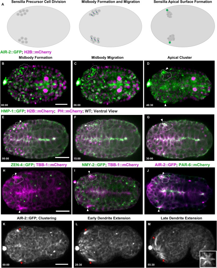
Fig. 7.
Midbody components label dendrites of sensilla neurons. (A) Diagram of SPC divisions with Aurora B (green; midbody ring in magenta). (B-D) Cytokinesis in SPCs expressing AIR-2::GFP (green; H2B::Cherry in magenta) gives rise to multiple midbodies (dashed outline, B,C) that cluster together (arrowheads, D). (E-G) HMP-1::GFP accumulates at the furrow and midbody (arrowheads, E,F), accumulates at the apical cluster, and remains at the tip (G) during dendrite extension. (H) ZEN-4::GFP (green; microtubules in magenta) is internalized and degraded before the microtubule-rich cluster forms (arrowheads). (I) NMY-2::GFP (green; microtubules in magenta) remains at the tip of the dendrite as it extends (arrowheads). (J) PAR-6::mCherry (green) and AIR-2::GFP (magenta) colocalizes to the cluster (arrowheads), indicating that this is the apical surface. (K-M) AIR-2::GFP labels the dendrites during extension. Inset in M is a rotated maximum z-projection of sensilla after dendrite extension. Time shown in minutes:seconds. Scale bars: 10 μm.

