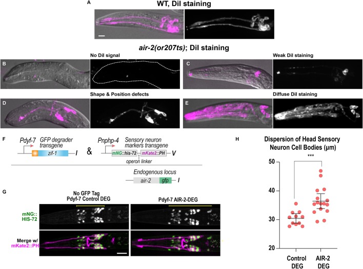
Fig. 8.
Cytokinesis mutants have disrupted sensilla neuron morphology. (A-E) Dendrite and neuron morphology revealed by DiI staining in L1 larvae. (A) In wild type (WT), two dendrite bundles and amphid and phasmid neurons are labeled. (B-E) air-2(or207ts) mutants show no DiI signal (B), weak signal (C), dendrite shape and positioning defects (D) and diffuse staining throughout the head of the animal (E). Dashed line outlines indicate animal position in B. (F) Construct used for post-mitotic degradation of AIR-2::GFP in sensory neurons. (G) Sensory neuron nuclei (green) and plasma membranes (magenta) for the indicated conditions. Scale bars: 10 μm. (H) Quantification of sensory neuron cell body distribution, measured as indicated by the yellow lines in G. Error bars are the 95% confidence interval. ***P<0.001 (two-tailed unpaired t-tests in GraphPad Prism; n=12 for control DEG and n=16 for AIR-2 DEG).

