Fig. 6.
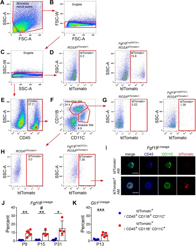
Alveolar macrophages phagocytose particles from Fgf18CreERT2-labeled cells. (A-H) Fgf18CreERT2 ROSAtdTomato/+ mice were injected with Tam daily from P5 to P8 and collected on P9, P15 or P31 for fluorescence-activated cell sorting. Sorted cells were gated against (A) debris and (B,C) doublets. (D) Gating strategy to identify tdTomato+ cells, set on control ROSAtdTomato/+, mice showing enrichment in Fgf18CreERT2/+; ROSAtdTomato/+ mice. (E-H) Sequential gating strategy to identify populations of tdTomato+ interstitial macrophages (CD45+ CD11B+ CD11C−) and alveolar macrophages (CD45+ CD11B− CD11C+). (G,H; left) Fluorescence minus one (FMO) control stained with all antibodies but without the tdTomato fluorophore were used as a negative control. All representative plots were generated from P9 sorted cells except FMOs, which were generated from P31 sorted cells. (I) Image of pooled flow-sorted cells from P9 tdTomato− and tdTomato+ alveolar macrophages. (J,K) Quantification of the percentage of interstitial macrophages (CD45+ CD11B+ CD11C−) and alveolar macrophages (CD45+ CD11B− CD11C+) that were gated as tdTomato+ in (J) Fgf18CreERT2/+; ROSAtdTomato/+ mice and (K) Gli1CreERT2/+; ROSAtdTomato/+ mice induced from P5 to P8 and collected on P13. Student's t-test, *P<0.05; **P<0.01; ***P<0.001. Mann–Whitney, #u=0.029. n=5 (P9, P13), n=4 (P15, P31). Scale bars: 10 µm. Data are mean±s.d. MΦ, macrophage.

