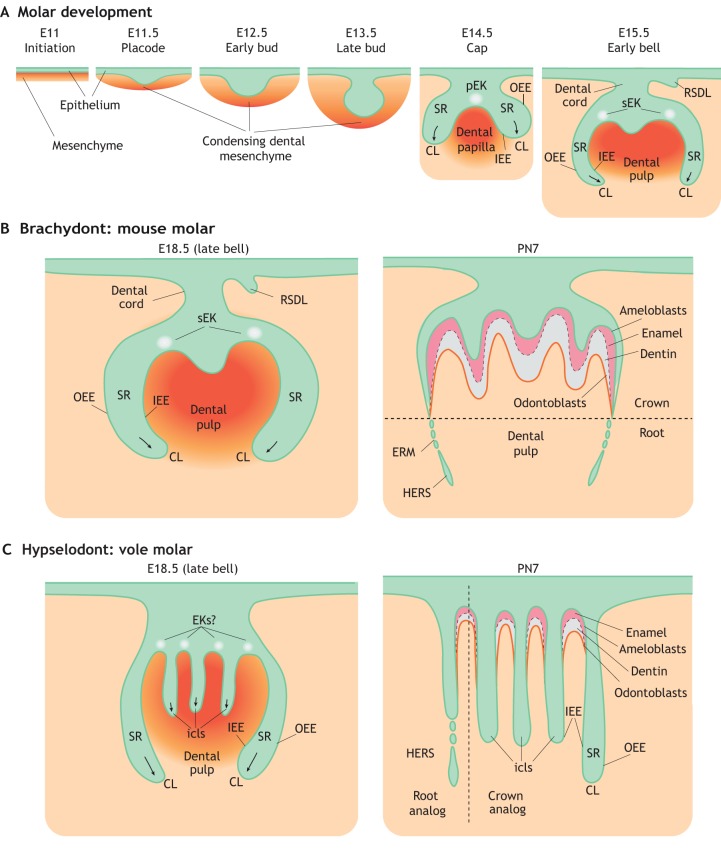
Fig. 1.
Schematic of rodent molar development. (A) The initiation of mouse molar formation starts a little later than E11 with the thickening of dental epithelium (green). The tooth bud then forms via invagination of the dental epithelium into the underlying condensing mesenchyme (orange) at E12.5-E13.5. Signals sent from the primary (pEK) and secondary (sEK) enamel knots guide the formation and elongation of the cervical loop (CL), with the inner (IEE) and outer (OEE) enamel epithelium surrounding the stellate reticulum (SR). The rudimentary successional dental lamina (RSDL), which is a transient structure in most species, also begins to form at this time. (B) During the later stages of mouse molar development, the dental epithelium of the CL grows apically to become a transient structure called Hertwig's epithelial root sheath (HERS) and the epithelial cell rests of Malassez (ERM), facilitating root formation. (C) During the later stages of molar development in voles, multiple intercuspal loops (icls) form and persist throughout the postnatal stages. These are then responsible for enamel deposition in between the icls. PN, postnatal day.

