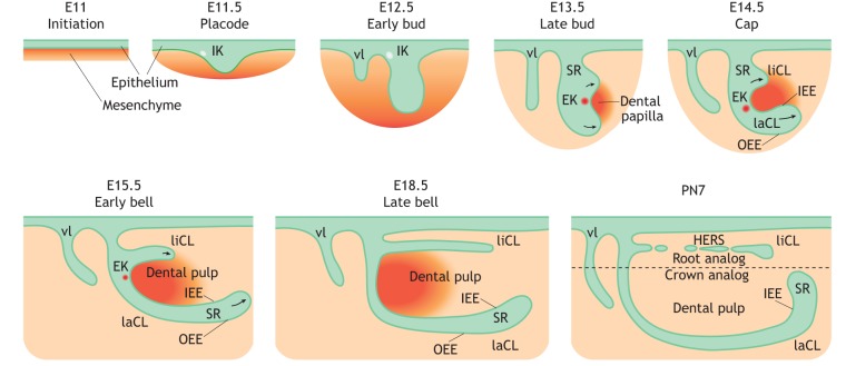
Fig. 2.
Schematic of mouse incisor development. The development of the mouse incisor initiates at around E11. Dental epithelium (green) invaginates into the mesenchyme (orange), forming the dental placode at around E11.5. A population of non-mitotic cells within the placode accumulates to form an early signaling center termed the initiation knot (IK), which guides the formation of the tooth bud. Vestibular lamina (vl) forms adjacent to the developing tooth bud. During the bud-to-cap transition, the signaling activity of the IK switches to the enamel knot (EK). At the cap stage at around E14.5, lingual and labial cervical loops (liCL and laCL) form on each side of the EK in an asymmetrical pattern along the labial-lingual axis around the dental papilla. Continuous folding further divides dental epithelium into inner (IEE) and outer (OEE) enamel epithelium, surrounding the stellate reticulum (SR) region. The development of the laCL proceeds through the bell stage, whereas the formation of the liCL is disproportionally slower on the medial side. Hertwig's epithelial root sheath (HERS) then forms at the lingual side. PN, postnatal day.

