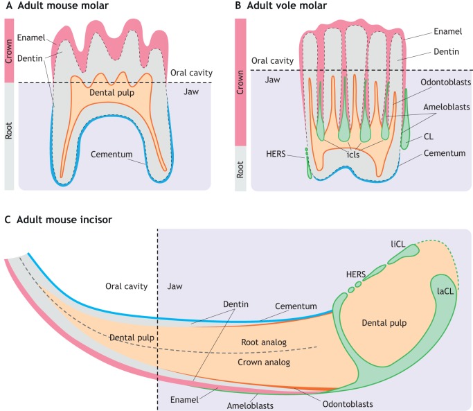
Fig. 3.
Schematic of adult rodent tooth structure. (A) In the adult mouse molar, crown eruption results in the loss of dental epithelial tissue. Thus, enamel produced by epithelial-derived ameloblasts is only deposited on the surface of the crown part of the molar. Mesenchymal-derived odontoblasts produce dentin around the dental pulp, and cementum mineralization covers the root portion of the molar. (B) In the vole molar, intercuspal loops (icls) persist throughout the adult stages, allowing the continuous growth of the crown. In addition, a larger proportion of the crown is buried inside the jaw bones compared with the mouse molar. Both Hertwig's epithelial root sheath (HERS) and the cervical loop (CLs) are preserved in adult vole molars. (C) In the adult mouse incisor, lingual and labial cervical loops (liCL and laCL) persist throughout the adult stages. Only the laCL contains dental stem cells, allowing a continuous supply of enamel-producing ameloblasts at the labial crown analog of mouse incisors. Dental pulp is enclosed by dentin, produced by mesenchyme-derived odontoblasts. The lingual root analog is covered by cementum, which helps the incisor anchor to the jaw bone.

