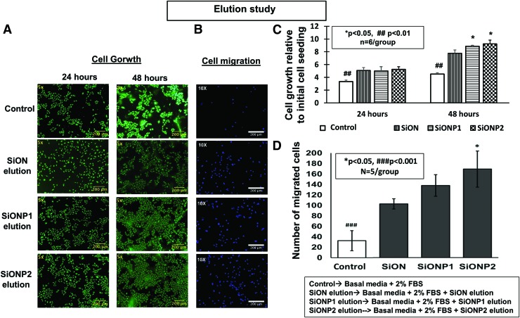
FIG. 3.
(A) Effect of elution from amorphous silica PECVD-coating scaffolds on HUVEC proliferation. Pictures after Calcein-AM staining (24 and 48 h). Scale bar = 200 μm (B) Effect of elution from PECVD-coating implants on HUVECs membrane transwell migration (24 hours). (B) Fluorescent pictures of nuclei fluorescent staining. Scale bar = 200 μm. (C, D) Bar graphs show the number of growth and migrated cells. ANOVA, *p < 0.05, ##p < 0.01, ###p < 0.001 indicate statistical significance. PECVD, plasma-enhanced chemical vapor deposition. Color images are available online.

