FIG. 1.
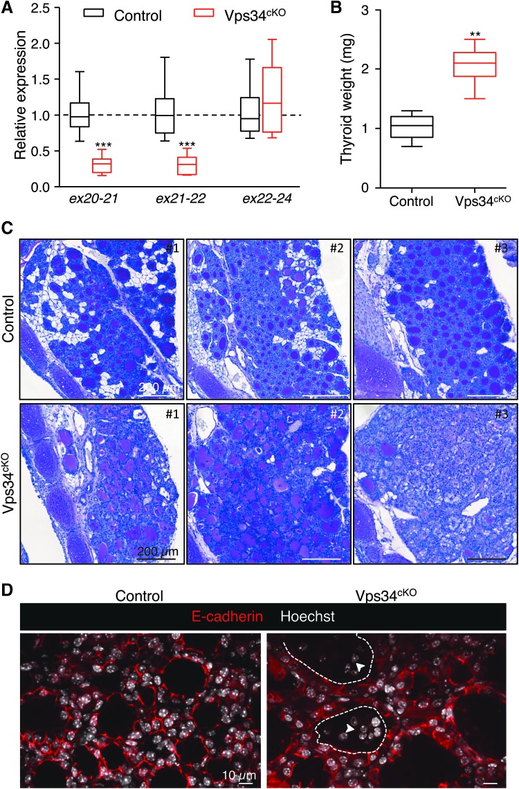
Genetic and histopathological characterization. (A) Extensive genetic excision of Vps34 exon 21. Compared with control (black boxes), Vps34cKO thyroid (red boxes) shows ∼70% reduction in exon 21 mRNA level at P14. The unchanged mRNA level spanning exons 22–24 serves as control. Boxes with median and percentiles of 10 WT and 11 cKO samples; ***p < 0.0001 by Mann–Whitney nonparametric test. (B) Increased thyroid weight. Compared with control (black boxes), Vps34cKO thyroid (red boxes) shows a twofold increase in thyroid weight. Boxes with median and percentiles of 8 WT and 6 cKO samples; **p < 0.01 by Mann–Whitney nonparametric test. (C) Histopathological evidence for colloid exhaustion. As compared with control thyroid tissues (n = 3) where all follicles show regular lumen filling with homogeneous and intense PAS staining, Vps34cKO thyroids (n = 3) present fewer, mostly centrally located, PAS-stained follicles and with weaker staining intensity, and other follicles that appear empty (for further quantification; Supplementary Fig. S1). (D) Vps34cKO thyrocytes are altered and follicles contain abundant nonepithelial cells. Nuclei are labeled by Hoechst (shown in white); thyrocyte basolateral contours are labeled for E-cadherin (red); two lumen boundaries are delineated by broken lines. As compared with control thyroid follicles, Vps34cKO follicular structures are frequently irregular and contain additional cells inside the lumen (arrowheads). These cells are not labeled for E-cadherin. cKO, conditional knockout; mRNA, messenger RNA; PAS, Periodic-Acid–Shiff; WT, wild type.

