Figure 8.

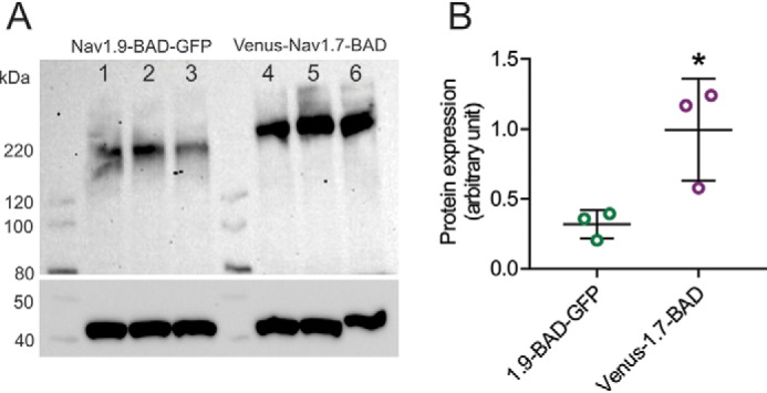
Steady-state protein levels of Nav1.9-BAD-GFP and Venus-Nav1.7-BAD in transiently transfected HEK293 cells. A, Western blotting of cell lysate of HEK293 cells transfected with either Nav1.9-BAD-GFP or Venus-Nav1.7-BAD. The blot was probed with pan-sodium channel (top) or β-actin (bottom) antibodies. Three independent transfections of different batches of HEK293 cells were analyzed: lanes 1–3 (Nav1.9-BAD-GFP) and lanes 4–6 (Venus-Nav1.7-BAD). B, protein expression levels were quantified using ImageLab software and presented as mean ± S.D. (error bars). A 3.1-fold difference was observed in protein expression between Nav1.9-BAD-GFP (0.32 ± 0.10 AU; n = 3) and Venus-Nav1.7-BAD constructs (1.00 ± 0.36 AU; n = 3), p < 0.05. Significance was determined using a two-tailed unpaired t test.
