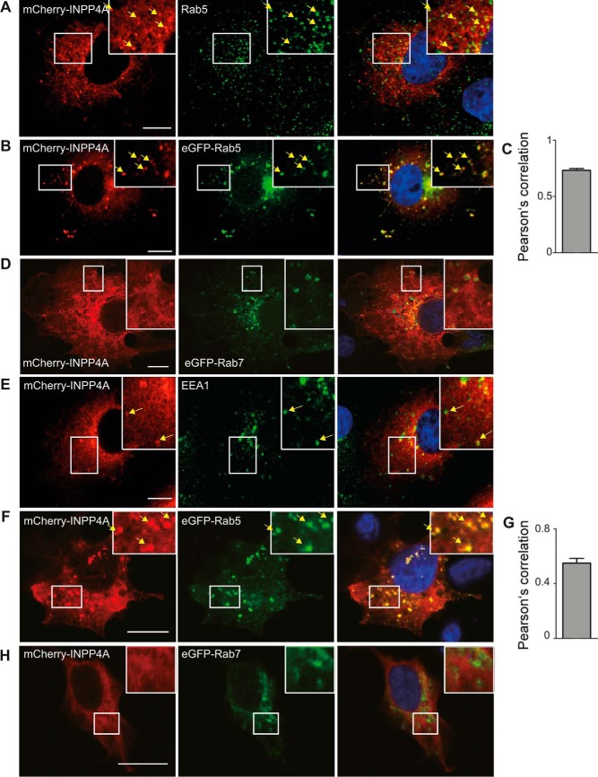
Figure 4.
INPP4A partially localizes to EEA1- and Rab5-containing endosomes. A, B, D, and E, confocal images of COS7 cells transiently expressing mCherry-INPP4A immunostained for endogenous Rab5 (A), overexpressed eGFP-Rab5 (B), overexpressed eGFP-Rab7 (D), or endogenous EEA1 (E). Note the partial colocalization of mCherry-INPP4A with the early endosome markers Rab5 and EEA1. Scale bars = 10 μm. C and G, Pearson's correlation coefficients determined from two-color confocal microscopy imaging of COS7 (C) and HAP1 (G) cells expressing mCherry-INPP4A and eGFP-Rab5. Mean ± S.E.; 61 and 46 cells were analyzed. F and H, confocal images of HAP1 cells transiently expressing mCherry-INPP4A and eGFP-Rab5 (F) or eGFP-Rab7 (H). Scale bars = 10 μm.

