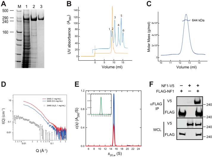
Figure 1.
Full-length neurofibromin is a high-affinity dimer. A, SDS-PAGE analysis of purified full-length neurofibromin. Lanes are molecular mass markers (M) with the sizes of relevant bands shown in kDa, clarified extract from the insect cell expression (lane 1), elution pool from IMAC chromatography (lane 2), and purified protein after size-exclusion chromatography (lane 3). B, analytical SEC trace of purified neurofibromin (orange) compared with molecular weight standards (blue). Standards used were blue dextran (peak 1), thyroglobulin (peak 2, 669 kDa), ferritin (peak 3, 440 kDa), aldolase (peak 4, 158 kDa), conalbumin (peak 5, 75 kDa), and ovalbumin (peak 6, 43 kDa). C, SEC-MALS analysis of full-length neurofibromin. D, SAXS/SANS analysis of full-length neurofibromin. Red (1 mg/ml) and blue (0.5 mg/ml) lines are SAXS data from runs with two different concentrations of neurofibromin, and open circles represent SANS data using 1 mg/ml neurofibromin. E, sedimentation velocity absorbance c(s) profiles for NF1 at 0.6 μm (red) and 1.2 μm (blue) based on data collected at 280 nm. The inset shows the corresponding c(s) profile for NF1 at 25 nm (green) based on absorbance data collected at 230 nm. F, Western blotting of immunoprecipitation of differentially epitope-tagged NF1 proteins from HEK293 cells. The top two gel sections contain lysate purified with anti-FLAG antibodies. The bottom two sections contain WCL. In both cases, the samples are probed with antibodies to the FLAG or V5 epitopes as noted. Molecular mass of standards is noted on the right in kilodaltons.

