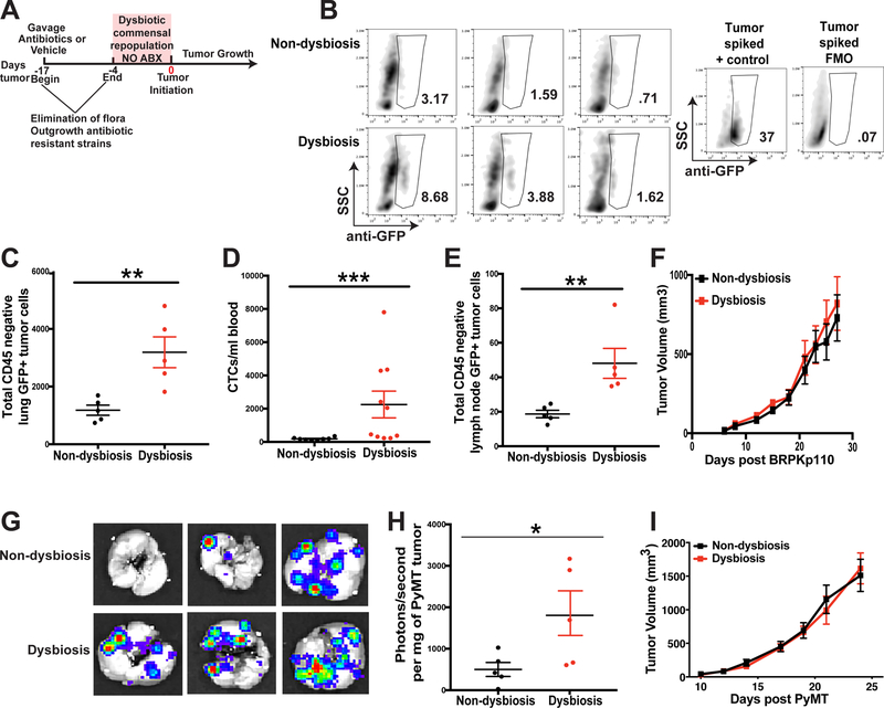
Figure 1. Commensal dysbiosis enhances mammary tumor cell dissemination.
A. Experimental design for antibiotic-induced dysbiosis and tumor initiation. C57BL/6 mice were orally gavaged for 14 days with a broad-spectrum cocktail of antibiotics or an equal volume of water as a vehicle control. Gavage was ceased four days prior to tumor initiation in the fourth mammary fat pad. Tumor size was measured by calipers every 2–3 days after reaching a palpable size. B. Representative plots demonstrating GFP+ tumor cell quantitation in the lungs. The anti-GFP gate was chosen based upon fluorescence minus one (FMO) controls and a stained lung sample spiked with BRPKp110 tumor cells. Numbers represent percent cells within the anti-GFP gate of total live cells. C-F. C57BL/6 mice were treated as described in Fig. 1A. 27 days after BRPKp110 tumor initiation, GFP+ tumor cell dissemination was quantified in lung tissue (C), peripheral blood (D), and tumor-draining axillary lymph nodes (E) by flow cytometry. Data is represented as absolute number of GFP+CD45− cells of live, singlet cells. F. Growth kinetics of BRPKp110 mammary tumors. Representative of at least three independent experiments with 5 mice/group. G-I. C57BL/6 mice were treated as described in Fig. 1A. 25 days after PyMT-luciferase tumor initiation, tumor cell dissemination was quantified in lung tissue by bioluminescence. G. Representative images of bioluminescence in lungs from advanced tumor-bearing mice bearing PyMT-luciferase tumors. H. Quantitation of luminescence represented as photons/second normalized to final tumor burden for mice with PyMT tumors. I. Growth kinetics of PyMT mammary tumors. Representative of two independent experiments with 5 mice/group.

