. 2020 Jan 3;34(1):143–153. doi: 10.21873/invivo.11755

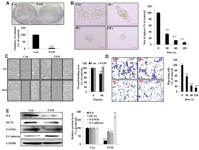
Figure 2. Plasma-activated medium (PAM) suppresses the malignancy of Aspc1 cells. A: The day after cancer cells had attached to the bottom of plates and the medium was removed and PAM added. Upper panel: The colony-forming ability in Aspc1 cells exposed to PAM (16.4 kV-120 s) for 7 days as detected by colony-formation assay; lower panel: quantification of colony-formation data. B: The sphere-forming ability of Aspc1 cells exposed to PAM (16.4 kV for 30 s, 60 s and 120 s) for 7 days as determined by sphere formation assay (Scale bar: 100 μm). Right panel: quantification of size of colonies. C: Wound-healing assay. Left: image; right: quantification of wound healing area. D: Transwell assay Left: image; right: quantification of cell migration ability (scale bar: 100 μm). E: Levels of interleukin 6 (IL6), octamer-binding transcription factor 4 (OCT4), NANOG, and E-cadherin proteins in Aspc1 cells after 6-h treatment with PAM (16.4 kV-120 s) as measured using western blot. The data are presented as the mean±SD of three independent experiments. Significantly different at *p<0.05; **p<0.01; ***p<0.001 vs. the control (Con).

Figure 2