. 2020 Jan 3;34(1):143–153. doi: 10.21873/invivo.11755

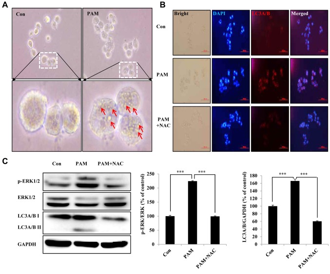
Figure 5. Plasma-activated medium (PAM) induces autophagic cell death of Aspc1 cells by reactive oxygen species (ROS). A: Imaging of morphology of Aspc1 cells exposed to PAM (16.4 kV-120 s) for 6 h (scale bar: 200 μm). Arrows: Microbubble structures. B: Immunocytochemistry assay of autophagy-associated protein light chain 3 alpha/beta (LC3A/B) in Aspc1 cells treated with PAM (16.4 kV-120 s) with and without N-acetyl cysteine (NAC) for 6 h (scale bar: 100 μm). C: Western blot analysis was performed to check the expression of p-extracellular-regulated kinase (ERK), p- ERK and LC3A/B in Aspc1 cells treated with PAM (16.4 kV-120 s) with and without NAC for 6 h. GAPDH: glyceraldehyde 3-phosphate dehydrogenase. The data are presented as the mean±SD of three independent experiments. ***Significantly different at p<0.001 vs. the control (Con).

Figure 5