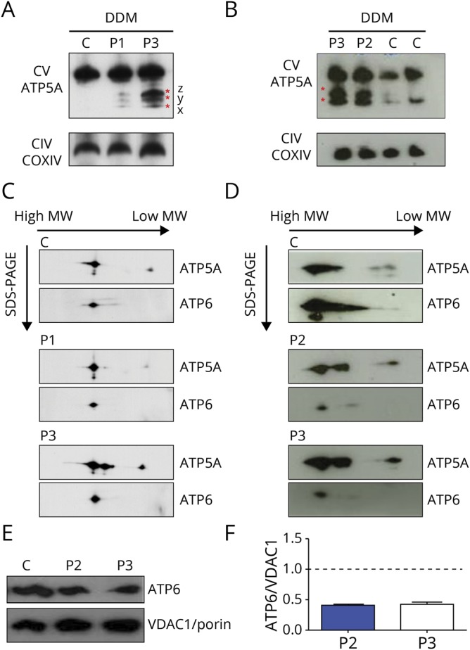
Figure 2. One-dimensional and 2-dimensional BNGE.

(A and B) Immunovisualization of complex V in 1-dimensional BNGE of enriched mitochondria fractions extracted from fibroblasts and muscles and solubilized with DDM. Control (C), patient 1 (P1), patient 2 (P2), and patient 3 (P3) are shown. Three F1 x, y, and z subcomplexes are present in fibroblasts of P1 and P3, whereas only 2 of 3 subassemblies are present in the muscle of P3 and P2. See discussion for details. Anti-ATP5A and anti-COXIV used to visualize complex V and complex IV, respectively. (C and D) Denaturing 2-dimensional BNGE of enriched mitochondria fractions extracted from fibroblasts and muscle and solubilized with DDM confirmed the presence of ATP synthase subcomplexes in P1, P2, and P3. Residual ATP6 protein is found incorporated into the fully assembled complex V in P1, P2, and P3. (E) Western blot of muscle samples show reduced ATP6 protein in patients (P2, P3) compared with the control. (F) Densitometry analysis of (E) performed in 2 different experiments. Values are normalized to controls. Error bars represent SEM. ATP = adenosine triphosphate; BNGE = blue-native gel electrophoresis; CIV = complex IV; COXIV = cytochrome c oxidase IV; CTR = control; CV = complex V; DDM = n-dodecyl-β-d-maltoside; MW = molecular weight; SDS-PAGE = sodium dodecyl sulfate-polyacrylamide gel electrophoresis; SEM = standard error of the mean; VDAC1 = voltage dependent anion channel 1.
