. 2019 Aug 5;55(69):10214–10217. doi: 10.1039/c9cc04145a

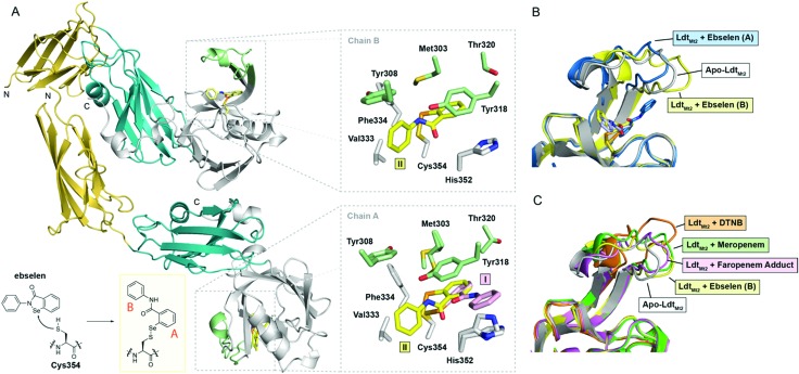
Fig. 2. Crystallographic studies of LdtMt2 with ebselen. (A) View from the crystallographically observed structure of LdtMt2 in complex with ebselen. The two immunoglobulin-related domains are in yellow and blue, while the catalytic domain is in white; the active-site loop region (lid) of the catalytic domain (residues 300–323) is in green. The inset shows the expected complex formed from ebselen and Cys354. Also shown are views of the active sites of chains A and B, with sticks coloured according to the cartoon representation, highlighting the two ebselen conformations observed (I, II). (B) Structural alignment of chains A and B of the complex derived from ebselen and LdtMt2 (blue and yellow cartoons, respectively) with the apo-enzyme (white cartoon), highlighting variations in the active site lid. (C) Overlay of LdtMt2 complex structures, showing variations in the active site lid. The unmodified enzyme (white cartoon) and ebselen adduct (chain B; yellow cartoon) structures are overlaid with LdtMt2 complexes derived from 5,5′-dithiobis-(2-nitrobenzoic acid) (DTNB; orange cartoon; PDB ; 5LB1),10 meropenem (green cartoon; PDB ; 3VYP),11 and faropenem (which fragments to form a 3-hydroxybutyryl group; pink cartoon; PDB ; 5LBG).10.

Fig. 2