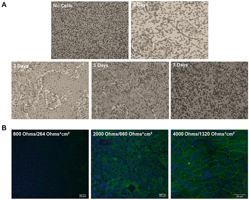
Fig. 6.
Human lung epithelial Calu-3 cell confluence over a single week’s culture in 10% FBS imaged using an inverted microscope with 10× magnification (a). Monolayer formation hides Transwell insert pores throughout the culture duration as seen from the initial image at time zero (top left) to the completion of monolayer formation at the conclusion of a week (bottom right). Confocal microscopy images of ZO-1 (green) and DAPI (blue) stained junctions and nuclei, respectively, display monolayer formation as a function of increased TEER resistance (b). Reproduced from [57]. https:/creativecommons.org/licenses/by-nc-nd/4.0/

