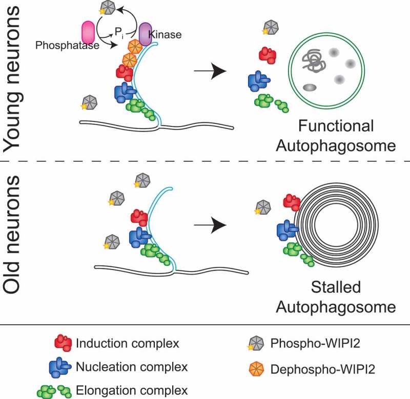
Figure 1.

Model of neuronal autophagosome biogenesis changes during aging. In neurons from young adult mice (top panel), dephosphorylation of WIPI2B at the phagophore allows autophagosome biogenesis to continue, ultimately yielding a stereotypical autophagosome, marked with LC3B (green membranes), enclosing bulk cytoplasm. Autophagosome biogenesis complexes are as differently colored protein complexes (see legend at bottom of figure). In neurons from aged mice (lower panel), localized dephosphorylation of WIPI2B does not occur, resulting in autophagic multilamellar vesicles that retain the components of the autophagosome biogenesis complexes, but do not recruit LC3B. These multilamellar structures do not appear to contain bulk cytoplasm.
