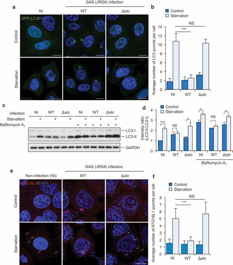
Figure 1.

GAS inhibits starvation-induced autophagy in a SLO-dependent mechanism. (a and b) Starvation-induced LC3-puncta formation in GAS-infected or non-infected (NI) cells. Confocal micrographs (a) and quantification (b) of LC3-puncta formation in HeLa cells stably expressing GFP-LC3 infected with GAS JRS4 strain wild-type or isogenic Δslo mutant for 2 h, and subsequently incubated with regular (control) or starvation medium for 1 h. Cells were fixed, and cellular and bacterial DNA was stained with DAPI. Scale bars: 10 μm. (c and d) HeLa infected with indicated GAS strains were incubated with starvation medium with or without bafilomycin A1 and analyzed by immunoblotting with indicated antibodies (c). Intensity ratio of LC3-II:LC3-I were normalized to that in non-infected (NI) cells without bafilomycin A1 (d). (e and f) Starvation-induced phagophore formation in GAS-infected or non-infected cells. Confocal micrographs (e) and quantification (f) of ATG16L1-puncta formation in HeLa cells infected with GAS JRS4 WT or Δslo mutant for 2 h, and subsequently incubated with regular (control) or starvation medium for 1 h. Cells were fixed, and stained with anti-ATG16L1 antibody. Scale bars: 10 μm. Data in (b, d, and f) are mean ± SEM of 3 independent experiments. Data were tested by two-tailed Student’s t-test: ***P < 0.001. NS, not significant.
