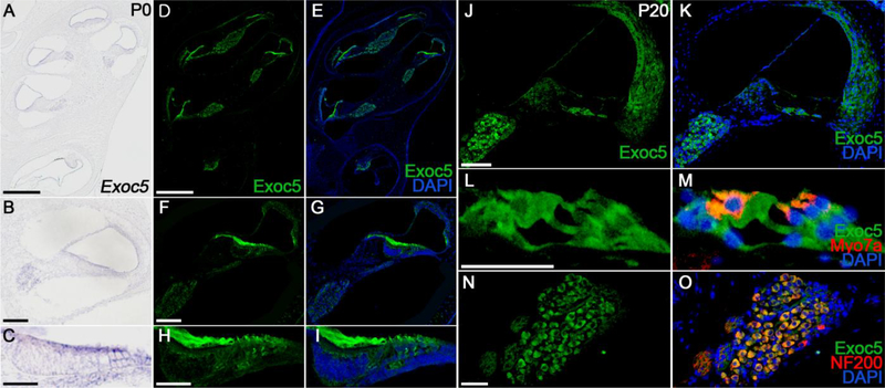
Fig. 1. Spatial pattern of EXOC5 expression in the inner ear.
(A-I) Expression pattern of EXOC5 was examined by in situ hybridization (A-C) and immunohistochemistry (D-I) at P0. The mRNA (violet) and protein (green) expression levels of EXOC5 were compared between the whole inner ear (A, D, and E), middle turn of the cochlea (B, F, and G) and the organ of Corti (C, H, and I). (J-O) EXOC5 immunohistochemistry was performed in the middle turn of the cochlea at P20. EXOC5 expression (green) was observed in both the Organ of Corti (L and M) and spiral ganglion (N and O). Hair cells and spiral ganglions were labeled with Myo7a (M, red) and NF200 (O, red), respectively. Nuclei were counterstained with DAPI (blue). Scale bars, 200 μm in (A, D, E), 50 μm in (B, F, G, J, K), 20 μm in (C, H, I), and 25 μm in (L-O).

