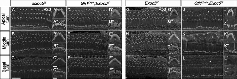
Fig. 4. Ultrastructural evaluation of the organ of Corti and stereocilia of hair cells in Gfi1Cre/+;Exoc5f/f mice.
The ultrastructure of stereociliary bundles from Exoc5f/f and Gfi1Cre/+;Exoc5f/f mice was analyzed using SEM at P20 (A-F, A′-F′, and A″-F″) and P50 (G-L, G′-L′, and G″-L″). (A-L) The Organ of Corti. (A′-L′) High magnification of an outer hair cell (OHC). (A″- L″) High magnification of an inner hair cell (IHC). Abnormal arrangement of rows of OHCs are indicated by asterisks. Scale bars, 15 μm in (A-L) and 2 μm in (A′-L′ and A″-L″).

