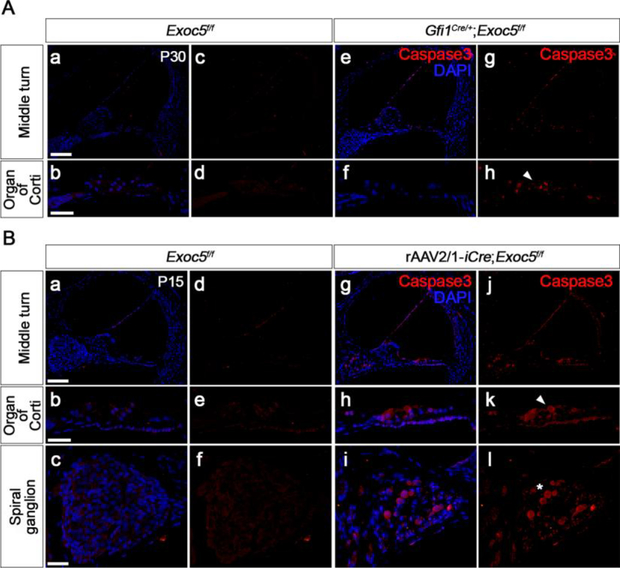
Fig. 9. Apoptosis in the sensory epithelium of Gfi1Cre/+;Exoc5f/f and rAAV2/1-iCre;Exoc5f/f mice.
Apoptosis was examined by Caspase3 immunostaining in the inner ear of Gfi1Cre/+;Exoc5f/f mice at P30 (A) and rAAV2/1-iCre;Exoc5f/f mice at P15 (B). Activated Caspase3-positive cells were labeled with Alexa Fluor® 555. Caspase3 activation in hair cells and spiral ganglions are indicated by arrow heads and asterisks, respectively. Nuclei were counterstained with DAPI (blue). Scale bars, 100 μm in (middle turn panels) and 25 μm in (organ of Corti and spiral ganglion panels).

