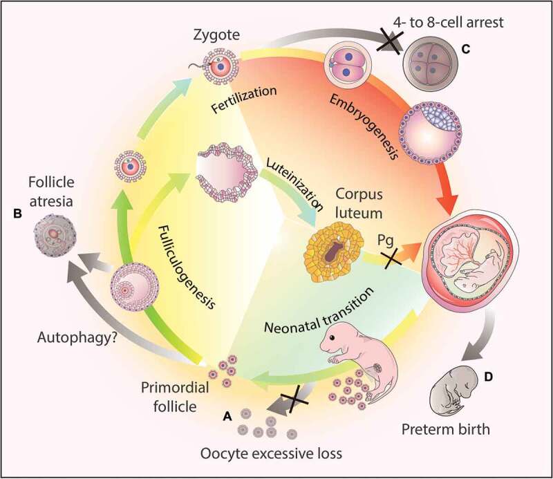
Figure 3.

Autophagy-dependent resource allocation in the female reproductive processes. (a) Autophagy might protect the oocytes from excesive loss in the neonatal ovaries. (b) Autophagy participates in follicle atresia, albeit the exact mechanism remains to be elucidated. (c) Autophagy actively participates in early embryogenesis, and the disruption of the autophagic flux results in 4- to 8-cell arrest. (d) Hormone synthesis. After ovulation, the corpus luteum forms from the remains of the ovulated follicles to secrete progesterone (Pg). Once autophagy is disrupted in this process, the synthesis of progesterone decreases due to insufficient lipid droplets in the luteal cells, and finally leads to preterm birth.
