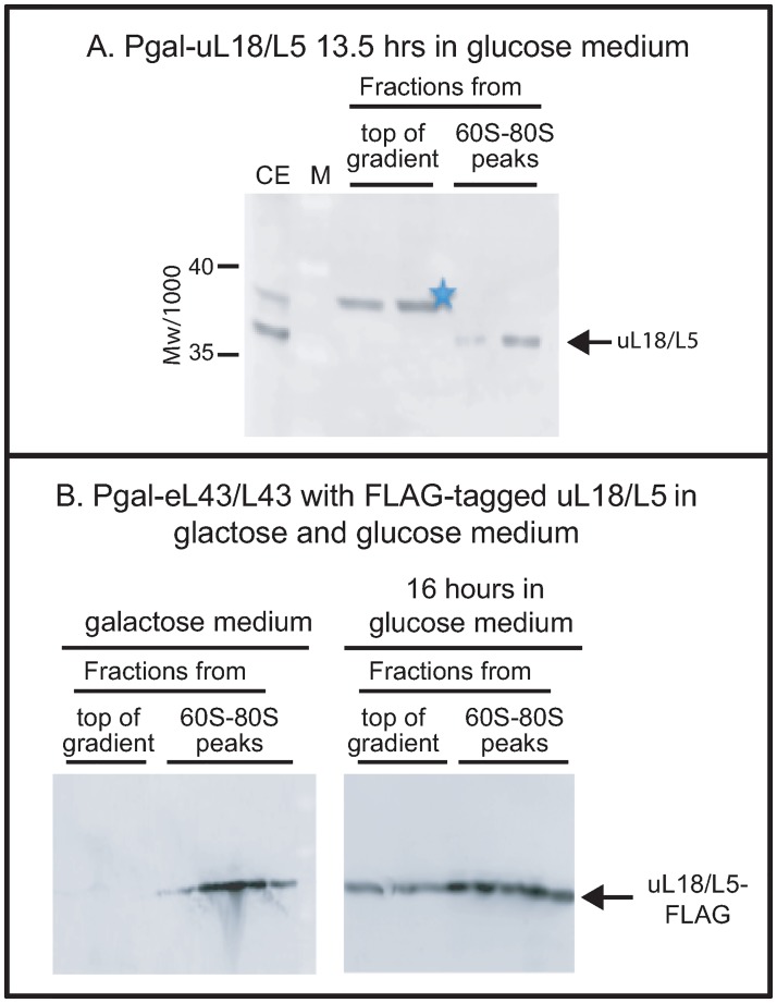
Fig 1. Analysis of the specificity of anti-uL18/L5.
(A) The uL18/L5 reactive band seen close to the top of the sucrose gradient after repressing eL43/L43 or eL40/L40 formation (Figs 2 and 3) is absent after repressing uL18/L5 synthesis. Pgal-uL18/L5 was grown in galactose medium and shifted to glucose medium. A lysate prepared after repression of uL18/L5 gene for 13.5 hours was fractionated on a sucrose gradient and consecutive fractions from the top of the gradient and the 60S-80S ribosome peaks were analyzed by western blot stained with anti-uL18/L5. (B) Distribution of FLAG-tagged uL18/L5 (uL18/L5-FLAG) in sucrose gradients loaded with lysates prepared before and after repressing eL43/L43 synthesis. Pgal-eL43/L43 was transformed with a plasmid harboring a constitutively expressed gene for uL18/L5-FLAG. The resulting strain was grown in galactose medium and shifted to glucose medium for 16 hours. Lysates prepared from cells before and after the shift were fractionated on sucrose gradient and aliquots of consecutive fractions from the top of the gradient and the 60S-80S peaks were analyzed for content of FLAG-tagged protein by western blot. The western blots in this figure were not cropped. M: Molecular weight markers/1000. CE: Crude cell Extract.

