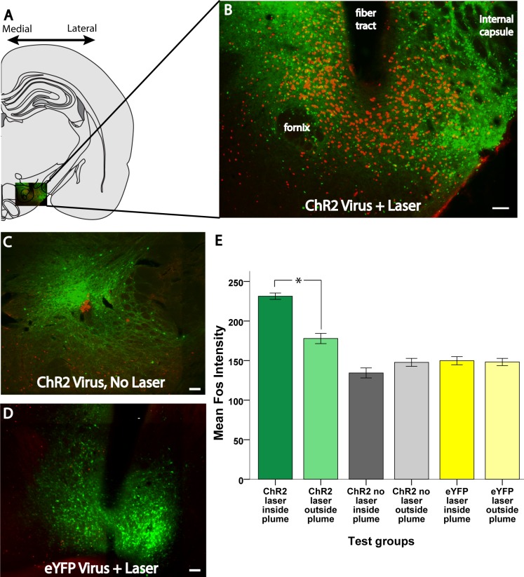
Fig 2. Examples of virus expression (green) and Fos-like immunoreactivity (red) in different treatment conditions.
A: Hemisection of a coronal map modified from a rat brain atlas (29). B: an inset of the lateral hypothalamus expanded out, giving an example of a robust ChR2 laser-induced Fos plume. C, D: Examples of virus and Fos expression in animals that received ChR2 virus without laser stimulation and eYFP virus with laser stimulation. E. Average intensity of Fos granules inside vs outside plumes for each experimental group and condition. Scale bars represent 100 μm unless otherwise stated. *: p < 0.05.

