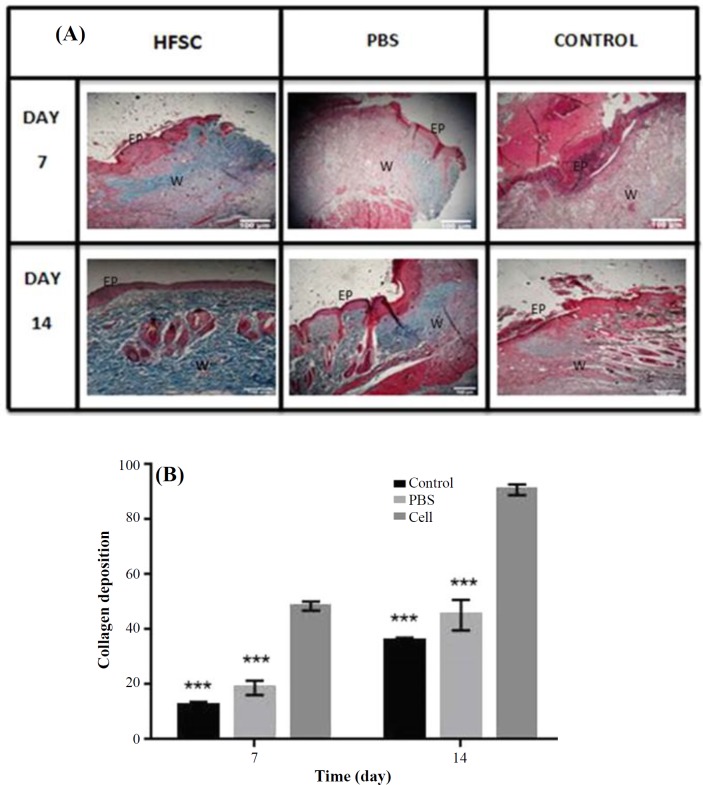
Fig. 7.
Histological analysis of burn wound healing in rat skin with Masson’s trichorome stain. (A) The processed tissue was assessed microscopically after Masson’s trichorome in HFSCs, PBS, and control groups at 7 and 14 days post burn; (B) Histological analysis of collagen deposition on days 7 and 14 after wound induction (n = 6; analysis of variance, mean ± SD; ***p < 0.001); EP, epidermis; W, wound bed

