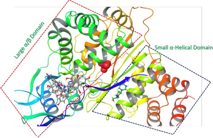
Fig 7.
Ribbon representation of TP (PDB:4LHM): It is showing a large α/β domain (red dotted lines) and a small α-helical domain (blue dotted lines). The inhibitor AZZ (green ball and stick) occupies the nucleoside binding pocket of TP while the phosphate binding site is in the α/β domain of TP. Compounds 3, 9, 14, 22, 27, and 29 (grey ball and stick) binds in the α/β domain of TP.

