
Figure 5.
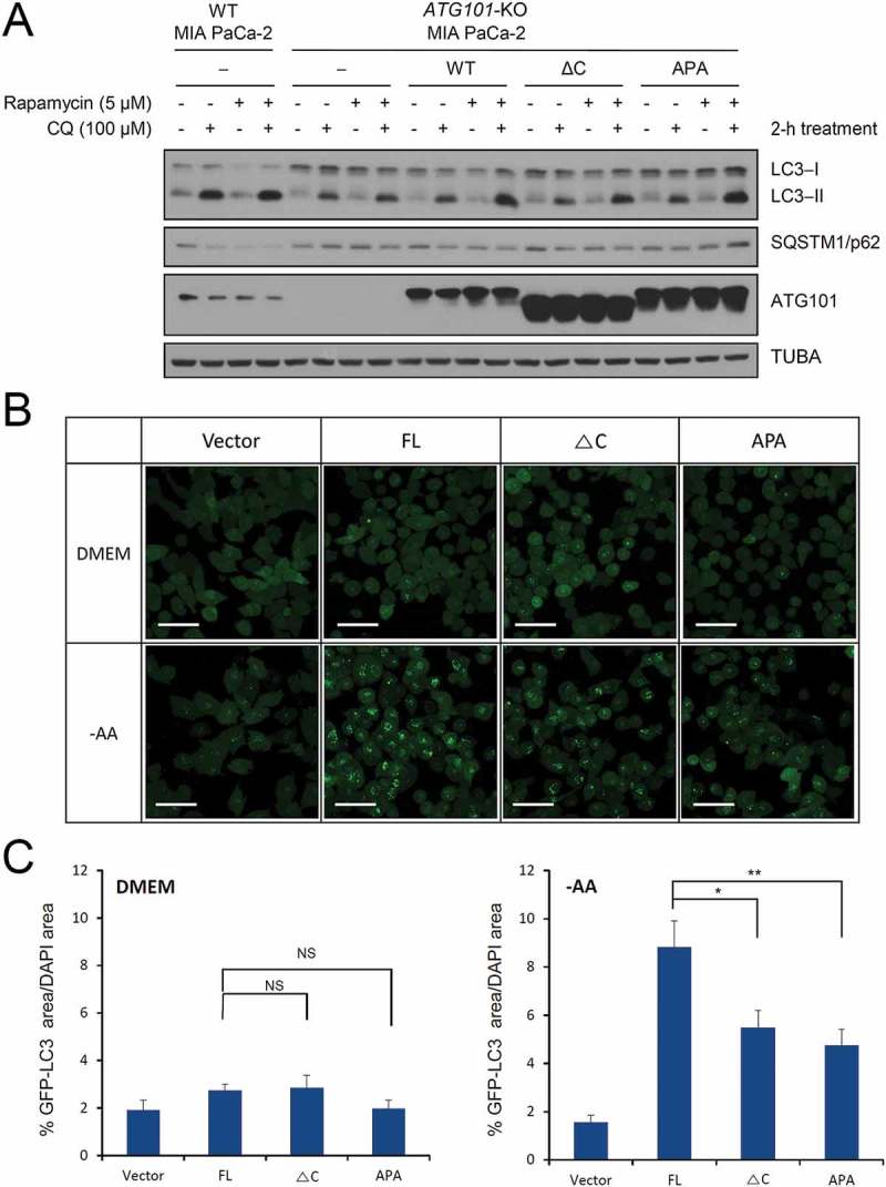
The role of the C-terminal region of ATG101 in the autophagy pathway. Autophagy defect shown in ATG101 knockout cells is restored by the expression of full-length ATG101 but not ATG101ΔC. (a) ATG101-knockout MIA PaCa-2 cells were transfected by a full-length wild-type of ATG101 (WT), a C-terminal deletion mutant of ATG101 (ΔC), and full-length ATG101 APA mutant (APA), respectively, and incubated for 48 h. After replacement with complete media containing 5 μM rapamycin (Sigma, R8781) with or without 100 μM chloroquine (CQ; Sigma, C6628), cells were further incubated for 2 h. For western blotting, cells were harvested and immunoblotted for ATG101, LC3, SQSTM1, and TUBA. (b) GFP-LC3 puncta were analyzed by live imaging using fluorescence microscopy after 2 h incubation of either complete (DMEM) or amino acid-depleted (-AA) media. Scale bar: 50 μm. (c) Quantification data for GFP-LC3 puncta area are expressed as a percentage of the DAPI area within the cell. NS, *, and ** mean P > 0.05, P < 0.05, and P < 0.01, respectively.