
Figure 4.
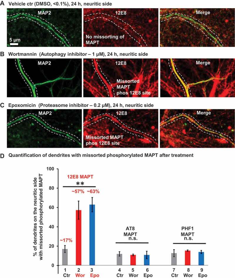
Missorting of MAPT into dendrites coincides with elevated phosphorylation at the 12E8 epitope, but not at the AT8 or PHF1 sites. Rat hippocampal neurons (DIV 21–25) cultured in microfluidic chambers treated on the neuritic side for 24 h either with DMSO (control, a), or with the autophagy inhibitor wortmannin (b) or proteasomal inhibitor epoxomicin (c). Phosphorylation-dependent MAPT antibody 12E8 was used to probe the phosphorylation state of MAPT at S262/S356 residues and MAP2 antibody was used to highlight dendrites. Images of the dendrites on the neuritic side are shown highlighting a dendrite with or without phospho-MAPT. (a-c) In vehicle-treated control (DMSO, < 0.1%) MAPT sorts mainly to the axons (a). Treatment with wortmannin (1 µM, 24 h) or epoxomicin (0.2 µM, 24 h) on the neuritic side causes an increase of MAPT, in the dendrites, which is phosphorylated at the 12E8 site (b and c). Scale bar: 5 µm. (d) Quantification of dendrites on the neuritic side showing colocalization of MAP2 with the phospho-MAPT antibodies 12E8 (n > 700 dendrites/dendritic branches from 5 experiments; one-way ANOVA with Tukey’s post hoc test; F [2,12] = 12; **p < 0.01), AT8 (n = > 70 dendrites/dendritic branches from 3 experiments; one-way ANOVA with Tukey’s post hoc test; F [2,6] = 0.05; p = 0.09) or PHF1 (n = > 30 dendrites/dendritic branches 3 experiments; one-way ANOVA with Tukey’s post hoc test; F [2,6] = 0.31; p = 0.74). ns, not significant.