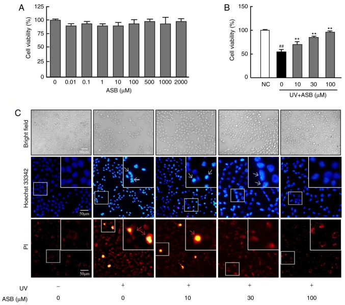
Figure 1.
Protective effect of ASB on UV-induced HaCaT cells. (A) The cells were treated with 0, 0.01, 0.1, 1, 10, 100, 500, 1,000 and 2,000 µM ASB for 24 h. The cell viability was determined by MTT assay (n=5 for each group). (B) The cells were preincubated with ASB (10, 30 and 100 µM) and irradiated with UV (90 mJ/cm2). Cell viability was measured by MTT assay (n=5 for each group). (C) The cells were preincubated with ASB (10, 30 and 100 µM) and irradiated with UV (90 mJ/cm2). Cell apoptosis and death were measured by staining with Hoechst 33342 and PI. The blue arrowheads indicate apoptotic cells, and the red arrowheads indicate dead cells. The images were examined by bright field and fluorescence microscopy. The results are expressed as the mean ± SD. ##P<0.01 vs. NC group; **P<0.01 vs. UV-alone group. UV, ultraviolet; ASB, andrographolide sodium bisulfite; NC, normal control; PI, propidium iodide.

