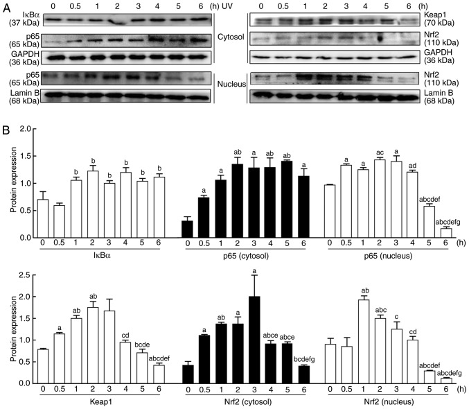
Figure 3.
Changes in the Nrf2 and NF-κB signaling pathways in HaCaT cells after UV irradiation. (A) Nuclear and cytoplasmic proteins were extracted from the cultured cells at 0, 0.5, 1, 2, 3, 4, 5 and 6 h after UV irradiation (90 mJ/cm2). The protein expression levels of NF-κB-mediated p65, and IκBα and Nrf2-mediated Nrf2 and keap1, were measured by western blotting. (B) Relative changes in protein intensities were quantified by densitometric analysis and are presented as bar diagrams (n=3 for each group). The results are expressed as the mean ± SD. aP<0.05 vs. the 0 h group; bP<0.05 vs. the 0.5 h group; cP<0.05 vs. the 1 h group; dP<0.05 vs. the 2 h group; eP<0.05 vs. the 3 h group; fP<0.05 vs. the 4 h group; gP<0.05 vs. the 5 h group. UV, ultraviolet; IκBα, NF-κB inhibitor-α; keap1, kelch-like ECH-associated protein 1; Nrf2, nuclear factor E2-related factor 2.

