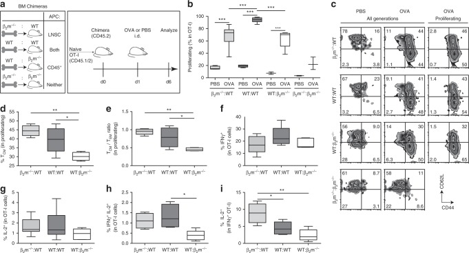
Fig. 1. CD8+ T cells educated by lymph node stromal cells display memory-like characteristics.
a Experimental schematic, highlighting nomenclature for BM chimeras according to MHCI-competent cell types (left), and timeline (right) for transfer of CFSE-labeled OT-I cells (i.v.), OVA challenge (50 μg; i.d.), and sacrifice. Skin-draining LNs were analyzed by flow cytometry. b Quantification of cell proliferation via CFSE dilution on transferred OT-I cells. c Representative contour plots of CD44 and CD62L expression gated on all transferred cells (left, center) or on proliferating transferred cells (right). d, e Phenotype of proliferating, transferred cells quantified according to d frequency of TCM (CD44+CD62L+) and e the ratio of TCM (CD44+CD62L+) to Teff/EM (CD44+CD62L−). f Frequency of intracellular IFNγ+, g IL-2+, and h bifunctional IFNγ+ IL-2+ cells among all transferred cells after 5 h ex vivo re-stimulation with SIINFEKL followed by intracellular staining. i Frequency of IL-2+ cells within IFNγ+ OT-I cells. Representative data pooled from two independent experiments (n = 3–5 each). Whiskers: Min to Max, median c, e, f; *p ≤ 0.05, **p ≤ 0.01, ***p ≤ 0.001 by one-way ANOVA followed by Bonferroni post-test.

