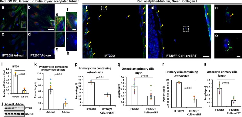
Fig. 4. Deletion of IFT20 in osteoblasts cause cilia loss.
Micrographs representing the primary osteoblasts either infected with Ad-null Adenovirus (a, c, e, g) as a control or infected with Ad-cre Adenovirus (b, d, f, h) to achieve IFT20 deletion under serum starvation condition for 48 h (a–h). IFT20 transcriptional expression levels (i) and translational expression levels (j) were validated to be down-regulated under the Ad-cre viral treatments. The percentage of ciliated cells was quantified and was significantly reduced in IFT20 deletion (n > 3) (k). Micrographs representing Z-stacked 3D-deconvolution processed images captured from the cells aligned on cortical bone of postnatal day 11 IFT20f/f and IFT20f/f;Col1-creERT femur using Leica DMI6000 inverted epifluorescence microscope under 40X lens (l–o). Red fluorescent signals detected immunofluorescent staining of Acetylated-tubulin and green fluorescent probed Collagen I and DAPI was depicted in blue. Quantitative comparisons of the primary cilia containing cells percentage (n = 3) (p) and the primary cilia length (n = 27) (q) in osteoblast. Quantitative comparisons of the primary cilia containing cells percentage (n = 3) (r) and the primary cilia length (n = 27) (s) in osteocyte. Scale bar (a, b, c, d, l, m) 20 μm, Scale bar (e, f, g, h, n, o) 5 μm.

